News & Research on Psychology | ShareYrHeart
The brain and gut are linked; find out how
Published
on
Summary: The brain and gut are linked. The new technology made by MIT engineers can study how hunger, mood, and different illnesses are controlled by the brain.
I am sorry, but there is no text provided for me to simplify. Please provide the text for me to assist with simplification. The main points of the text are in easy language.
Source: Massachusetts Institute of Technology
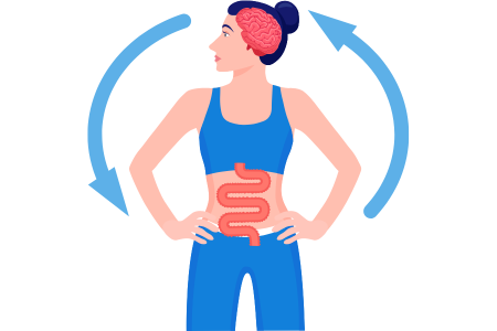
How the brain and stomach are connected
Engineers made a thing that can look at how the brain and stomach are connected. Researchers used tiny fibers with sensors and light to control the connections between the stomach and the brain in mice.
The brain and stomach talk to each other to help control eating and other things our body does. The big communication system we use can affect our thinking and might cause some brain problems.
MIT engineers created a new tool to explore links between things. Scientists have used tiny fibers with sensors and lights to control connections between the brains and guts of mice.
Scientists showed that they could make mice feel full or want rewards by changing cells in their gut. In their future research, they want to study the connections between gut health and brain conditions like autism and Parkinson’s disease.
This is cool because we can use technology to control how our stomach acts and how we eat. We can now study how our stomach and brain talk to each other in a really accurate way by using a scientific method called optogenetics. And we can study this in animals while they are moving around,” explains Polina Anikeeva, the Matoula S. Salapatas is someone who teaches about materials science and engineering. They also teach about the brain and cognitive sciences. Salapatas is part of MIT’s Research Laboratory of Electronics and is involved with MIT’s McGovern Institute for Brain Research.
Anikeeva wrote a new study that is being published in a science journal called Nature Biotechnology. The Salapatas are the people who wrote the paper Sahasrabudhe: Sahasrabudhe. Who studies at MIT; Laura Rupprecht, who is a researcher at Duke University; Sirma Orguc. Sio is a researcher at M.T.; and Tural Khudiyev, who used to study at MIT.
How your brain and body are connected
The McGovern Institute started something called the K last year. Lisa Yang Brain-Body Center is researching how the brain works with other organs in the body. The center does research on how interactions affect behavior and health. They want to develop treatments for different illnesses in the future.
Our body and brain always talk to each other in both directions, Anikeeva explains. We used to believe that the brain was like a boss that told the other parts of the body what to do. We’ve learned that there’s a lot of communication going back to the brain. Which might control some things we thought were only controlled by the brain.
Anikeeva wants to study the messages that go back and forth between the brain and the gut. Cells in the stomach and intestines affect how hungry or full we feel through signals that they send to the brain and chemicals that they release.
Understanding the effects of hormones on the brain has been hard because we can’t quickly and accurately measure the signals in the brain that occur very quickly.
To study the connection between the gut and the brain, we needed a precise device that didn’t exist before. This device would help us measure the effects on brain function and behavior with accuracy up to milliseconds. “We decided to create it,” says Sahasrabudhe, who was in charge of making the gut and brain probes.
Researchers made a gadget with bendable strings that can do a lot of things and can be put inside body parts they want to study. To make small threads, Sahasrabudhe used a special method called thermal drawing. These threads are very thin and can have devices added to them, like sensors or electrodes.
The filaments have small tools that give off light and can make cells work.
They also have tiny pipes for giving medicine. The fibers can be made to work better in different body parts. The scientists made strong fibers to go inside the brain. Scientists created softer, rubbery fibers that won’t harm the digestive organs, like the intestine, but can still withstand the tough conditions inside them.
Sahasrabudhe says that we need technology that can connect with organs and the brain and record signals accurately to understand how they work together. We need to make certain cells in diforgans, the body parts of mice, active so that we can study how they behave and figure out how they work.
The tiny threads can be controlled without touching them by using a device that can be placed on the animal’s body during testing. Orguc and Harrison created a remote control system that doesn’t need wires. They worked with two professors at MIT to make it.
The way people drive
They used fibers to send a signal to a part of the brain that gives off dopamine. When the mice received a lot of dopamine, they wanted to go back to the same place where they got it.
The scientists wanted to see if they could make people want rewards by affecting their stomachs. They made the animals want to go back to a certain place by using fibers in their stomachs to release sugar. Which caused the brain to release a chemical called dopamine.
The researchers found that they could make the body crave rewards by using light to stimulate nerve endings in the gut, which controls digestion. They did this instead of using a sweet substance called sucrose. They worked with people from Duke University to make this discovery.
We found that people still showed a preference for this place even though we didn’t use brain stimulation like before. We are only making the gut work and watching how it affects the brain from outside, says Anikeeva.
Sahasrabudhe worked together with a postdoc named Rupprecht in Professor Diego Bohorquez’s group at Duke to test how well the fibers could control someone’s feeding habits. They discovered that the equipment could activate cells that make a hormone called cholecystokinin. This hormone makes you feel full. When the hormone was released, the animals weren’t hungry anymore, even though they hadn’t eaten for a while. The scientists proved that they could make cells that control appetite work better by using a chemical called PYY. When we eat very rich food, our body naturally makes PYY to help us feel full, and the researchers found a way to make it work even better.
Conclusion
The scientists want to use this tool to learn more about how the brain and the gut work together in diseases that affect the nerves. For example, research has found that kids with autism have a higher chance of having problems with their stomachs than other kids. They also have something in common with people who have anxiety and irritable bowel syndrome through their genes.
We can start thinking about whether there is a link between the stomach and the brain. If there is, we may be able to use this connection to treat certain conditions. By working on the stomach without having to touch the brain directly.
Source : Massachusetts Institute of Technology
Image Source : Canva
Hi, I’m Aarti, My Psychoanalytical approach towards my clients is to empower them to better their lives through improving their relationship with themselves. I believe shame and guilt is a common barrier to change. I aim to guide my clients through re authoring their narratives where shame, guilt, and other problems have less power and take up less space.
Related Posts
News & Research on Psychology | ShareYrHeart
Depressed Individuals Maintain Focus on Negatives Even After Recovery, Study Finds
Published
1 month agoon
August 24, 2023Summary: Depressed Individuals Maintain Focus on Negatives Even After Recovery, a study finds. In contrast to people who have never suffered a major depressive episode, people who have recovered from one typically spend more time processing negative information and less time processing positive information, which increases the likelihood of a recurrence.
Source: American Psychological Association
Lead author Alainna Wen, PhD, a postdoctoral scholar at the Anxiety and Depression Research Center at the University of California, Los Angeles, said, “Our research indicates that individuals with a previous history of depression dedicate a significant amount of time to analyzing negative stimuli, such as expressions of sadness, in comparison to positive stimuli like displays of happiness. This contrast is more pronounced when compared to emotionally healthy individuals with no such history. This divergence in processing suggests a heightened susceptibility to potential future depressive episodes. This susceptibility arises from the inherent nature of depression, which involves an abundance of negative thoughts and emotions, coupled with a scarcity of positive ones.”
The Journal of Psychopathology and Clinical Science published the research.
One of the most prevalent mental illnesses in the US is major depression. The National Institute of Mental Health estimates that 21 million American people (8.4% of the country’s population) reported having had serious depression at least once in 2020. Major depression can hinder or limit a person’s capacity to do important life tasks. It is characterized by a minimum of two weeks of a gloomy mood or a lack of interest in or enjoyment from daily activities.
Wen reports that relapse rates for major depressive disorder are still high despite the availability of effective therapies for the condition. After their first major depressive episode, over 50% of people will go on to have several episodes, and they frequently relapse within two years of getting better. To enhance treatment and prevent recurrence, researchers need to gain a deeper understanding of the risk factors associated with major depressive illness.
Research on Depressed Individuals
Researchers performed a meta-analysis of 44 trials, comprising 2285 healthy controls and 2081 patients with a history of severe depressed disorder, for this publication. Every study looked at how quickly people responded to neutral, positive, or negative stimuli. In several instances, participants were requested to press a separate button to represent a happy, sad, or neutral human face. In other cases, participants responded to favorable, unfavorable, or neutral words.
Regardless of whether the stimuli were pleasant, neutral, or negative, healthy participants reacted to both emotional and non-emotional stimuli faster than those with a history of depression. However, compared to controls, individuals who had previously experienced major depressive disorder took longer to process unpleasant emotional cues. When compared to individuals in major depression remission, healthy controls showed a significant difference in the amount of time they spent processing positive versus negative emotional stimuli; however, this difference did not show up when comparing the amount of time spent processing positive versus neutral or negative versus neutral stimuli.
Conclusion
According to Wen, the results generally imply that people with recurrent major depressive disorder not only exhibit a stronger bias for focusing on negative information over positive or neutral information, but they also appear to be less able to manage the information they absorb than people in good health.
“The results of this study have implications for depressed people’s treatment,” Wen stated. Merely centering on diminishing the analysis of negative data might not be adequate to avert a relapse of depression. Alternatively, approaches that enhance the processing of positive information could prove advantageous for patients.”
Source: American Psychological Association
Image Source: Canva
News & Research on Psychology | ShareYrHeart
Obesity and Cognitive Function: How Liraglutide’s Impact Offers Hope
Published
1 month agoon
August 21, 2023Summary: Obesity affects how the body uses energy and decreases the sensitivity of cells to insulin. The term “anti-obesity drugs” is gaining increasing usage for the treatment of obesity, attracting significant attention, particularly in the USA. Researchers have recently established that decreased insulin sensitivity in obese individuals has an impact on the learning of sensory associations. Just one dose of the anti-obesity medication liraglutide might normalize these alterations and restore the underlying brain circuit function.
Source: Max Planck Institute for Biology of Aging
Liraglutide improves cognitive function in people suffering obesity
The brain needs to be able to create associations in order to govern our conduct. This entails, for instance, linking a neutral external stimulus with a result occurring after the stimulus (for instance, the hotplate flashes red—you could burn your hand). The brain gains knowledge of the implications of how we respond to the initial stimulus in this way. The foundation for creating brain connections and the source of the motivational power in stimuli is associative learning. The dopaminergic midbrain, a part of the brain, is largely in charge of controlling it. Since this area includes a large number of receptors for the body’s signalling molecules, including insulin, it can modify our behaviour to meet physiological requirements.
But what occurs when obesity reduces the body’s sensitivity to insulin? Does this alter our brain activity, capacity for association learning, and hence, behaviour? Now, scientists at the Max Planck Institute for Metabolism Research have examined how well the learning of associations functions in individuals with normal body weight (high insulin sensitivity, 30 volunteers) and in individuals with obesity (reduced insulin sensitivity, 24 volunteers), as well as whether the anti-obesity medication liraglutide affects this learning process.
The brain’s capacity to link sensory stimuli is diminished by low insulin sensitivity in people suffering from obesity.
During the study, participants were administered either an evening trial injection of the medication liraglutide or a placebo. A substance known as a GLP-1 agonist, liraglutide activates the GLP-1 receptor in the body, increasing insulin levels and causing satiety. Administered once a day and often utilized to treat type 2 diabetes and obesity, it was followed by a learning exercise the next morning for participants. This exercise enabled researchers to assess the efficacy of associative learning. The study revealed that the ability to link sensory stimuli was less pronounced in obese individuals compared to participants with normal weight. Furthermore, brain activity in the regions encoding this learning behavior experienced a decrease.
Participants with obesity no longer displayed these abnormalities after just one dosage of liraglutide, and there was no distinction in brain activity between participants with normal weight and obesity. In other words, the medication brought brain activity back to that of people who were of a normal weight.
Conclusion
“These discoveries are critically important. Here, we demonstrate how basic behaviours like associative learning are influenced by both internal metabolic state and exterior environmental factors. Therefore, whether a person is overweight or not also affects how their brain develops associations between sensory input and motivation. According to studies showing that these medications restore a normal feeling of satiety, causing people to eat less and consequently lose weight, the normalisation we achieved with the drug in obese subjects fits with this finding, says study leader Marc Tittgemeyer from the Max Planck Institute for Metabolism Research.
Although it’s positive that current medications demonstrate beneficial effects on the brain activity of people with obesity, it’s worrisome that alterations in brain function can emerge even among younger individuals with obesity who do not have any other underlying medical issues. Future healthcare reforms should place considerably more emphasis on preventing obesity. According to Ruth Hanßen, MD, first author of the study and a physician at the University Hospital of Cologne, lifelong medication is the less desirable alternative in comparison to primary prevention of obesity and associated consequences.
Source: Max Planck Institute for Biology of Aging
Image Source: Canva
News & Research on Psychology | ShareYrHeart
Chronic Stress Reveals Distinct Responses in Brain Cells of Males and Females
Published
1 month agoon
August 18, 2023Summary: Chronic Stress Reveals Distinct Responses in Brain Cells of Males and Females. Researchers have shown that a subgroup of brain cells reacts to stress in males and females very differently. The results may help us understand the relationship between chronic stress and diseases including anxiety, depression, obesity, and diabetes and open the door to more specialized treatments for these ailments.
Source: Weizmann Institute of Science
Chronic stress results in an ongoing rise in mental and physical problems
Chronic stress results in an ongoing rise in mental and physical problems, which has a huge negative impact on society. Although not always in the same way, they have an impact on both men and women. Men and women handle stress in different ways, according to a wealth of information, but the reasons why these disparities exist are still unknown. In any case, there are no personalized treatments available for men or women. But Chen’s laboratory, which focuses on researching how the body reacts to stress, made the claim that cutting-edge research techniques might help alter the situation.
However, researchers obtained those results using research techniques that could mask significant variations in the reactions of specific cells or even entirely eliminate the functions carried out by relatively uncommon cells. Earlier investigations in other labs had discovered some sex differences in the response to stress. In contrast, Chen’s group employs cutting-edge techniques that enable researchers to examine brain activity at a previously unheard-of granularity—on the level of the individual cell—and may, as a result, throw new light on the disparities between the sexes.
The study’s principal investigator, According to Dr. Elena Brivio
The study’s principal investigator, According to Dr. Elena Brivio, “We focused on the area of the brain that serves as a main hub of the stress response in mammals, the paraventricular nucleus (PVN) of the hypothalamus, using the most sensitive research lens imaginable. We were able to map the stress response in male and female mice along three main axes by sequencing the RNA molecules in that part of the brain at the cellular level: how each type of brain cell in that region reacts to stress, how each type of cell previously exposed to chronic stress reacts to a fresh stress encounter, and how these responses vary between males and females.
The researchers mapped out gene expression for more than 35,000 individual cells, generating an extensive dataset that portrays an unparalleled picture of the stress response and underscores the differences in how males and females experience and react to stress.The researchers decided to publish the entire detailed mapping as part of the study and in accordance with open-access scientific principles. The website went live simultaneously with the publication of the study, providing other researchers with straightforward and convenient access to the data. In line with Brivio’s comments, the platform will enable researchers concentrating on a particular gene to observe how the gene’s activity alters in reaction to stress within a particular cell category, encompassing both males and females.
The thorough mapping has already enabled the researchers of Chronic Stress
The thorough mapping has already enabled the researchers to pinpoint numerous variations in gene expression, including those between males and females and between acute and chronic stress. The findings revealed, among other things, that different brain cells react to stress in males and females in various ways: Some cells are more sensitive to stress in males and some to stress in females. Researchers discovered that the oligodendrocyte, a type of glial cell crucial for supporting nerve cells and controlling brain activity, exhibited the most significant change within the brain area.
Dr. Juan Pablo Lopez emphasized that while neurons have taken the central spotlight in scientific research, they make up just approximately a third of the total population of brain cells. A former postdoctoral fellow in Chen’s group, he currently leads a research team at the Department of Neuroscience at the Karolinska Institute in Sweden. He emphasized that the method they employed enables them to achieve a more comprehensive and detailed understanding, encompassing every cell type and their interconnections within the specific brain region being examined.
Simple diversity by Chronic Stress
Clinical trials for new medications were only carried out on men up until the 1980s. The general consensus was that incorporating women in the study was superfluous and would merely complicate it by adding new variables like menstruation and hormonal shifts. Previously, researchers discouraged the utilization of female animals in preclinical experiments for similar reasons. Researchers formerly discouraged the use of female animals in preclinical experiments for the same reasons. There is no reason to believe that females would complicate the trials more than males, as it is now understood that male animals usually display greater molecular and behavioral variability than females.
Nevertheless, it’s a common practice in basic science to only use male subjects for tests. Our findings emphasize the significance of considering the gender aspect when it comes to stress-related health conditions, ranging from depression to diabetes. This is vital as it markedly influences the responses of diverse brain cells to stress,” remarks Chen. In the realm of neuroscience and behavioral science, Brivio further explains, “Even when a study doesn’t explicitly focus on gender distinctions, involving female subjects in the investigation remains crucial.” Equally crucial is the implementation of the most nuanced research methods, aiming to capture the most comprehensive portrayal of brain activity possible.
The research team was composed of Dr. Aron Kos, Stoyo Karamihalev, Andrea Ressle, Rainer Stoffel, and Dr. Mathias V. Schmidt, all affiliated with the Max Planck Institute of Psychiatry in Munich. Furthermore, the study involved Dr. Alessandro Francesco Ulivi from the Leibniz Institute for Neurobiology in Magdeburg, Germany, Dana Hirsch from Weizmann’s Veterinary Resources Department, and Dr. Gil Stelzer from Weizmann’s Life Sciences Core Facilities Department.
Source: Weizmann Institute of Science
Image Source: Canva
Trending